Gynaecology tests and symptom relief procedures
Information for patients under the care of the Gynae-Oncology Team
The hospital has contacted you to come for a biopsy or procedure. This leaflet will explain the following.
Who will be performing the tests, and where they will take place.
How to prepare for your appointment.
What to bring to your appointment.
What a biopsy is, and why you might need one.
What other procedures you may have.
We hope this leaflet answers some of the questions you may have. If you have any further questions or concerns, please speak to a member of your healthcare team.
Who will be performing the tests?
A specially trained doctor called a radiologist, will carry out each procedure listed in this leaflet.
Where will the tests take place?
The procedures usually take place in the Radiology Department, in one of the scan rooms.
In an emergency, some of the procedures may be performed on the ward.
How do I prepare for my appointment?
We may ask you to not eat or drink anything for a few hours before your biopsy. This is called fasting. We will include details of this in your appointment letter, or your clinical nurse specialist (CNS) will explain this to you.
Your doctor or CNS will tell you whether you can take your regular medications during your fasting period. They may give you other special instructions as well.
Do I need to bring anything to my appointment?
Please bring a list of your current medications with you to your appointment. Especially medications that affect blood clotting, such as aspirin or blood thinners.
Depending on which procedure you are having, you might be told to bring an overnight bag to your appointment.
To make sure all patients get the best care and attention during their procedure, your appointment may be delayed. We often arrange appointments at short notice, which means that the Radiology Department is not always able to honour the time slot given. It is extremely rare for anyone to have their procedure cancelled on the day for this reason. Please bring something to read or some light entertainment. Especially if you are likely to need 4 hour bed rest after your procedure.
Can I bring someone with me to hospital?
Yes, a relative or friend can come with you to hospital. However, they will not be able to stay with you for your procedure or wait on the ward. They can visit you on the ward after your procedure.
What is a biopsy and why might I need one?
During a biopsy, we take a small sample of cells or body tissue, usually with a needle. The skin around the biopsy site is numbed with a local anaesthetic, so it will not hurt. Afterwards, we examine the cells under a microscope.
Below is a list of the biopsies you may have. Before we ask for your consent to continue, we will explain what will happen during the biopsy. Please use the time before your procedure to ask the radiologist any last minute questions. Remember you can withdraw your consent for treatment at any time.
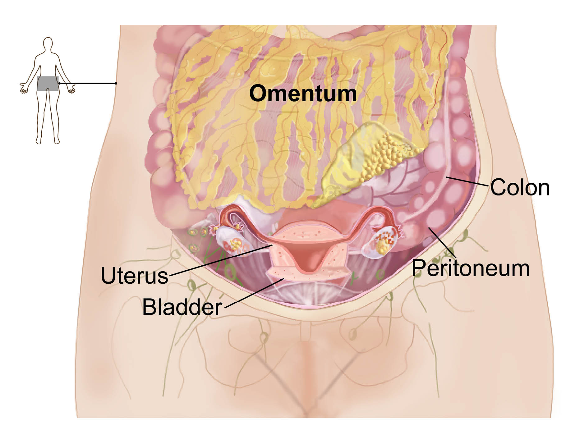
Omental biopsy
The omentum is a large fatty tissue layer that lines the organs in your abdomen. It is very often not the cause of your symptoms. However, it is close to most of the organs in your abdomen, so it tends to collect evidence of most conditions. It can help us diagnose your condition.
When there is evidence of deposits on your scans, we recommend a biopsy of the omentum as we can get to it easily. The radiologist uses an ultrasound scanner to find the right place to take the biopsy. This reduces the risk of injury to your surrounding organs.
During the biopsy, the radiologist takes a small sample of omental tissue with a needle. The skin around the biopsy site is numbed with a local anaesthetic, so this will not hurt. You need to stay in bed for the first 4 hours after this procedure. If you feel well after this period, you can go home.
Common risks affect between 1 in 10 and 1 in 100 people. These risks include tenderness and bruising after your procedure, but these will pass quickly.
Rare complications affect between 1 in 1000 and 1 in 10,000 people. These complications include infection, bleeding, and injury to internal organs.
Peritoneal biopsy
The peritoneum is the lining of the abdomen (see diagram above). It can sometimes collect evidence of conditions in the abdomen. We may recommend a biopsy of the peritoneum if there is evidence of deposits on your scans.
The radiologist uses an ultrasound or CT scanner during the procedure. This helps them find the right place to biopsy. It also reduces the risk of injury to your surrounding organs. After giving you a local anaesthetic to numb your skin, the radiologist uses a needle to take a small amount of the peritoneal tissue.
You need to stay in bed for the first 4 hours after this procedure. If you feel well after this period, you can go home.
Common risks affect between 1 in 10 and 1 in 100 people. These risks include tenderness and bruising after your procedure, but these will pass quickly.
Rare complications affect between 1 in 1000 and 1 in 10,000 people. These complications include infection, bleeding, and injury to internal organs.
Groin node biopsy
Lymph nodes (gland) are small round organs that are part of the body’s lymphatic system. They swell in response to infection, immune conditions, and cancer. Swollen nodes in the groin are easy to access for biopsy.
After giving the local anaesthetic to numb your skin, the radiologist uses a needle to take a sample of the cells from your lymph node. This is a short procedure. You can go home immediately afterwards.
Common risks affect between 1 in 10 and 1 in 100 people. These risks include tenderness, bruising, or bleeding.
What other procedures might I have?
Ascitic tap
During an ascitic tap the radiologist removes a small amount of ascitic fluid from your abdomen. Ascitic fluid is an abnormal fluid build-up of the abdomen. The radiologist numbs your skin with a local anaesthetic before removing the fluid using a needle.
An ultrasound scanner is used during the procedure, to find an area with a large amount of fluid. Using the scanner will reduce the risk of injury to your internal organs. The radiologist collects the cells in the fluid and sends them to a laboratory for examination.
You can go home shortly after your procedure.
It is normal to feel tender and have mild bruising around the area where the needle was inserted. This can last for a few days after your procedure. Ascitic fluid may also leak from the area of the sample in the first 72 hours.
Ascitic drain
The main aim of this procedure is to remove the excessive amounts of ascitic fluid you have in your abdomen. This should help relieve any tightness, swelling, and shortness of breath you are feeling. Ascitic fluid is an abnormal fluid build-up of the abdomen.
To do this, the radiologist places a drain in your abdomen. The drain removes as much ascitic fluid as possible. The radiologist numbs your abdomen with a local anaesthetic, before placing a fine tube into your abdomen.
The radiologist uses an ultrasound scanner during the procedure, to find an area with a large amount of fluid. Using the scanner will reduce the risk of injury to your internal organs. They collect the cells in the fluid and send them to a laboratory for examination.
For most people this is a day case procedure. However, in order to take as much fluid as possible, you might need an overnight stay in hospital to allow all the fluid to drain. Please pack an overnight bag in case this happens.
Common risks affect between 1 in 10 and 1 in 100 people. These risks include tenderness, mild bruising, a drop in blood pressure, and leakage of ascitic fluid from where the drain entered the body.
Rare complications affect between 1 in 1000 and 1 in 10,000 people. These are more serious and include an infection in your abdominal cavity and injury to internal organs.
Pleural drain
A pleural drain is when fluid gathers between the two layers of membrane covering your lungs. If this happens, the radiologist places a drain into your lungs to remove as much pleural fluid as possible. As a result, you will need an overnight stay in hospital to allow all the fluid to drain. Please pack an overnight bag if you are having this procedure.
A fine tube is placed in your chest. The radiologist uses an ultrasound scanner during the procedure, to find an area with a large amount of fluid. Using the scanner will reduce the risk of injury to your internal organs.
Common risks affect between 1 in 10 and 1 in 100 people. These risks include tenderness and bruising.
Rare complications affect between 1 in 1000 and 1 in 10,000 people. These are more serious and include injury to the lungs and shortness of breath. Shortness of breath is due to air getting into the pleural space (pneumothorax).
What happens next?
Once the laboratory has sent us your results, we will contact you by phone or letter to let you know what will happen next.
What if I feel unwell at home?
Contact the Gynaecological Assessment Unit, if when you get home:
you feel unwell; or
develop any of the common complications listed above for your procedure.
Go your nearest Emergency Department if you develop:
a temperature;
abdominal pain; or
breathing problems.
Contact details
If you have any questions or concerns either before or after your procedure, please contact one of the following.
Gynaecological Assessment Unit, Queen Elizabeth the Queen Mother (QEQM) Hospital, Margate
Telephone: 01843 235009Gynaecological Assessment Unit, William Harvey Hospital, Ashford
Telephone: 01233 651987
What do you think of this leaflet?
We welcome feedback, whether positive or negative, as it helps us to improve our care and services.
If you would like to give us feedback about this leaflet, please fill in our short online survey. Either scan the QR code below, or use the web link. We do not record your personal information, unless you provide contact details and would like to talk to us some more.
If you would rather talk to someone instead of filling in a survey, please call the Patient Voice Team.
Patient Voice Team
Telephone: 01227 868605
Email